
| Sequence Source Links | ||||||
|
||||||
| Database Satistics | |||
|
|||
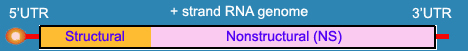
Structural Protein |
|
| E Protein (Major envelope protein E ) | |
| Description | The E-protein of dengue virus, a 60 KDa glycoprotein, which is embedded in lipid bilayer, may mediate both the virus attachment as well as penetration into cells. The structure of soluble E (sE) protein as elucidated by X-ray crystallography consists of three domains; domain I (DI), the N-terminal but structurally central domain, domain II (DII), the fusion domain containing the hydrophobic fusion peptide, and domain III (DIII), the putative receptor binding domain. One of these (domain II), an elongated, finger-like structure, bears a loop at its tip with a hydrophobic pocket lined by residues that influence the pH threshold for fusion. The pocket (residues 98−109 in dengue type 2), which accepts a hydrophobic ligand, opens and closes through a conformational shift in a beta-hairpin at the interface between two domains. These features point to a structural pathway for the fusion-activating transition and suggest a strategy for finding small-molecule inhibitors of dengue and other flaviviruses. The dimeric form of the E protein on viral membrane surface dissociates upon acidification, binds liposomes, and irreversibly trimerizes. These trimers cluster on the liposome surface and induce curvature which might promote fusion. The elongated trimer bears three "fusion loops" at one end, to insert into the host-cell membrane, and domains DI and DIII are at the other. |
| Function | Receptor binding and fusion with the host cell |
| Cellular Location | Anchored on the luminal side of the ER membrane |
| Assays | ELISAs to detect NS1 levels in solution have been published [Young et al. (2000), Alcon et al. (2002)]. |
| Structure | CRYSTAL STRUCTURE OF THE DENGUE 2 VIRUS ENVELOPE GLYCOPROTEIN IN THE POSTFUSION CONFORMATION |
References (Pubmed) |
14737159 , 12759475 |